×
Network disruptions
We have been experiencing disruptions on our local network which has affected the stability of these web pages.
We have been working with IT support team to get this fixed as a matter of urgency and apologise for any inconvenience.
PDB Information
| PDB | 5CA3 |
| Method | X-RAY DIFFRACTION |
| Host Organism | Escherichia coli BL21(DE3) |
| Gene Source | Escherichia coli (strain K12) |
| Primary Citation |
Crystal structure of the enzyme-product complex reveals sugar ring distortion during catalysis by family 63 inverting alpha-glycosidase.
Miyazaki, T., Nishikawa, A., Tonozuka, T.
J.Struct.Biol.
|
| Header | Hydrolase |
| Released | 2015-06-29 |
| Resolution | 1.800 |
| CATH Insert Date | 14 Aug, 2016 |
PDB Images (11)
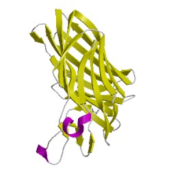
5ca3A01
CATH Domain 5ca3A01
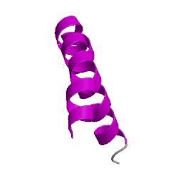
5ca3A02
CATH Domain 5ca3A02
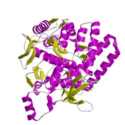
5ca3A03
CATH Domain 5ca3A03
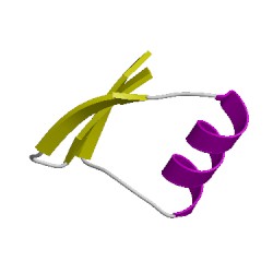
5ca3A04
CATH Domain 5ca3A04
5ca3B01
CATH Domain 5ca3B01
5ca3B02
CATH Domain 5ca3B02
5ca3B03
CATH Domain 5ca3B03
5ca3B04
CATH Domain 5ca3B04
PDB Chains (2)
| Chain ID | Date inserted into CATH | CATH Status |
| A | 15 Aug, 2016 | Chopped
|
| B | 15 Aug, 2016 | Chopped
|
CATH Domains (8)
UniProtKB Entries (2)
| Accession |
Gene ID |
Taxon |
Description |
| P42592 |
YGJK_ECOLI |
Escherichia coli K-12 |
Glucosidase YgjK |
| P42592 |
YGJK_ECOLI |
Escherichia coli K-12 |
Glucosidase YgjK |