×
Network disruptions
We have been experiencing disruptions on our local network which has affected the stability of these web pages.
We have been working with IT support team to get this fixed as a matter of urgency and apologise for any inconvenience.
PDB Information
| PDB | 3V3B |
| Method | X-RAY DIFFRACTION |
| Host Organism | Escherichia coli |
| Gene Source | Homo sapiens |
| Primary Citation |
Structure of the stapled p53 peptide bound to Mdm2.
Baek, S., Kutchukian, P.S., Verdine, G.L., Huber, R., Holak, T.A., Lee, K.W., Popowicz, G.M.
J.Am.Chem.Soc.
|
| Header | Ligase/Ligase Inhibitor |
| Released | 2011-12-13 |
| Resolution | 2.000 |
| CATH Insert Date | 22 Jan, 2012 |
PDB Images (7)
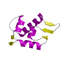
3v3bA00
CATH Domain 3v3bA00
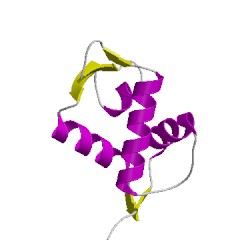
3v3bB00
CATH Domain 3v3bB00
PDB Chains (4)
CATH Domains (2)
UniProtKB Entries (2)
| Accession |
Gene ID |
Taxon |
Description |
| Q00987 |
MDM2_HUMAN |
Homo sapiens |
E3 ubiquitin-protein ligase Mdm2 |
| Q00987 |
MDM2_HUMAN |
Homo sapiens |
E3 ubiquitin-protein ligase Mdm2 |