×
Network disruptions
We have been experiencing disruptions on our local network which has affected the stability of these web pages.
We have been working with IT support team to get this fixed as a matter of urgency and apologise for any inconvenience.
PDB Information
| PDB | 3R4D |
| Method | X-RAY DIFFRACTION |
| Host Organism | Spodoptera frugiperda |
| Gene Source | Mus musculus |
| Primary Citation |
Crystal structure of mouse coronavirus receptor-binding domain complexed with its murine receptor.
Peng, G., Sun, D., Rajashankar, K.R., Qian, Z., Holmes, K.V., Li, F.
Proc.Natl.Acad.Sci.USA
|
| Header | Cell Adhesion/Viral Protein |
| Released | 2011-03-17 |
| Resolution | 3.100 |
| CATH Insert Date | 26 Jun, 2011 |
PDB Images (11)
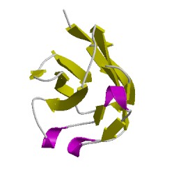
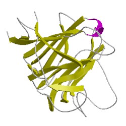
3r4dA01
CATH Domain 3r4dA01
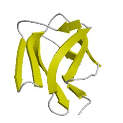
3r4dA02
CATH Domain 3r4dA02
3r4dB00
CATH Domain 3r4dB00

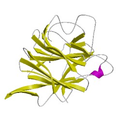
3r4dC01
CATH Domain 3r4dC01
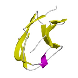
3r4dC02
CATH Domain 3r4dC02
3r4dD00
CATH Domain 3r4dD00
PDB Chains (4)
CATH Domains (6)
UniProtKB Entries (4)
| Accession |
Gene ID |
Taxon |
Description |
| Q9J3E7 |
Q9J3E7_9BETC |
Murine hepatitis virus |
Spike glycoprotein |
| P31809 |
CEAM1_MOUSE |
Mus musculus |
Carcinoembryonic antigen-related cell adhesion molecule 1 |
| Q9J3E7 |
Q9J3E7_9BETC |
Murine hepatitis virus |
Spike glycoprotein |
| P31809 |
CEAM1_MOUSE |
Mus musculus |
Carcinoembryonic antigen-related cell adhesion molecule 1 |