×
Network disruptions
We have been experiencing disruptions on our local network which has affected the stability of these web pages.
We have been working with IT support team to get this fixed as a matter of urgency and apologise for any inconvenience.
PDB Information
| PDB | 3KMD |
| Method | X-RAY DIFFRACTION |
| Host Organism | |
| Gene Source | |
| Primary Citation |
Crystal structure of the p53 core domain bound to a full consensus site as a self-assembled tetramer.
Chen, Y., Dey, R., Chen, L.
Structure
|
| Header | Dna Binding Protein/Dna |
| Released | 2009-11-10 |
| Resolution | 2.150 |
| CATH Insert Date | 25 Feb, 2010 |
PDB Images (11)
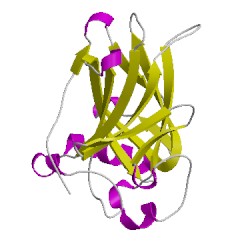
3kmdA00
CATH Domain 3kmdA00
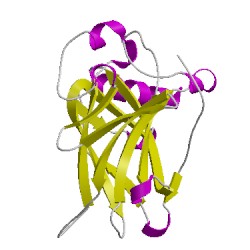
3kmdB00
CATH Domain 3kmdB00
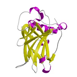
3kmdC00
CATH Domain 3kmdC00
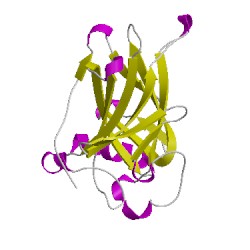
3kmdD00
CATH Domain 3kmdD00
PDB Chains (6)
CATH Domains (4)
UniProtKB Entries (4)
| Accession |
Gene ID |
Taxon |
Description |
| P04637 |
P53_HUMAN |
Homo sapiens |
Cellular tumor antigen p53 |
| P04637 |
P53_HUMAN |
Homo sapiens |
Cellular tumor antigen p53 |
| P04637 |
P53_HUMAN |
Homo sapiens |
Cellular tumor antigen p53 |
| P04637 |
P53_HUMAN |
Homo sapiens |
Cellular tumor antigen p53 |