×
Network disruptions
We have been experiencing disruptions on our local network which has affected the stability of these web pages.
We have been working with IT support team to get this fixed as a matter of urgency and apologise for any inconvenience.
PDB Information
| PDB | 2X76 |
| Method | X-RAY DIFFRACTION |
| Host Organism | |
| Gene Source | |
| Primary Citation |
The structure of PhaZ7 at atomic (1.2 A) resolution reveals details of the active site and suggests a substrate-binding mode.
Wakadkar, S., Hermawan, S., Jendrossek, D., Papageorgiou, A.C.
Acta Crystallogr. Sect. F Struct. Biol. Cryst. Commun.
|
| Header | Hydrolase |
| Released | 2010-02-24 |
| Resolution | 1.450 |
| CATH Insert Date | 13 Jun, 2010 |
PDB Images (3)
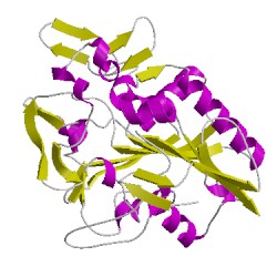
2x76A00
CATH Domain 2x76A00
PDB Chains (1)
| Chain ID | Date inserted into CATH | CATH Status |
| A | 13 Jun, 2010 | Chopped
|
CATH Domains (1)
UniProtKB Entries (1)
| Accession |
Gene ID |
Taxon |
Description |
| Q939Q9 |
Q939Q9_PAULE |
Paucimonas lemoignei |
PHB depolymerase PhaZ7 |