×
Network disruptions
We have been experiencing disruptions on our local network which has affected the stability of these web pages.
We have been working with IT support team to get this fixed as a matter of urgency and apologise for any inconvenience.
PDB Information
| PDB | 2V62 |
| Method | X-RAY DIFFRACTION |
| Host Organism | ESCHERICHIA COLI |
| Gene Source | HOMO SAPIENS |
| Primary Citation |
Structure of the Pseudokinase Vrk3 Reveals a Degraded Catalytic Site, a Highly Conserved Kinase Fold, and a Putative Regulatory Binding Site.
Scheeff, E.D., Eswaran, J., Bunkoczi, G., Knapp, S., Manning, G.
Structure
|
| Header | Transferase |
| Released | 2007-07-13 |
| Resolution | 1.700 |
| CATH Insert Date | 07 Nov, 2007 |
PDB Images (7)
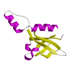
2v62A01
CATH Domain 2v62A01
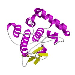
2v62A02
CATH Domain 2v62A02
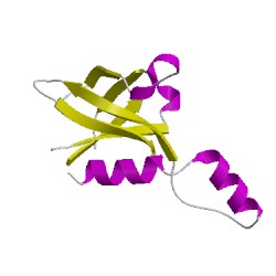
2v62B01
CATH Domain 2v62B01
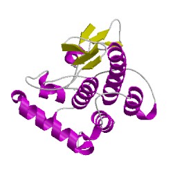
2v62B02
CATH Domain 2v62B02
PDB Chains (2)
| Chain ID | Date inserted into CATH | CATH Status |
| A | 08 Nov, 2007 | Chopped
|
| B | 08 Nov, 2007 | Chopped
|
CATH Domains (4)
UniProtKB Entries (2)
| Accession |
Gene ID |
Taxon |
Description |
| Q86Y07 |
VRK2_HUMAN |
Homo sapiens |
Serine/threonine-protein kinase VRK2 |
| Q86Y07 |
VRK2_HUMAN |
Homo sapiens |
Serine/threonine-protein kinase VRK2 |