×
Network disruptions
We have been experiencing disruptions on our local network which has affected the stability of these web pages.
We have been working with IT support team to get this fixed as a matter of urgency and apologise for any inconvenience.
PDB Information
| PDB | 2AT9 |
| Method | ELECTRON CRYSTALLOGRAPHY |
| Host Organism | |
| Gene Source | |
| Primary Citation |
The structure of bacteriorhodopsin at 3.0 A resolution based on electron crystallography: implication of the charge distribution.
Mitsuoka, K., Hirai, T., Murata, K., Miyazawa, A., Kidera, A., Kimura, Y., Fujiyoshi, Y.
J.Mol.Biol.
|
| Header | Photoreceptor |
| Released | 1998-12-17 |
| Resolution | 3.000 |
| CATH Insert Date | 05 Mar, 2006 |
PDB Images (3)
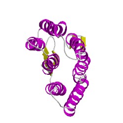
2at9A00
CATH Domain 2at9A00
PDB Chains (1)
| Chain ID | Date inserted into CATH | CATH Status |
| A | 05 Mar, 2006 | Chopped
|
CATH Domains (1)
UniProtKB Entries (1)
| Accession |
Gene ID |
Taxon |
Description |
| P02945 |
BACR_HALSA |
Halobacterium salinarum NRC-1 |
Bacteriorhodopsin |