×
Network disruptions
We have been experiencing disruptions on our local network which has affected the stability of these web pages.
We have been working with IT support team to get this fixed as a matter of urgency and apologise for any inconvenience.
PDB Information
| PDB | 1QZN |
| Method | X-RAY DIFFRACTION |
| Host Organism | Escherichia coli BL21 |
| Gene Source | Acetivibrio cellulolyticus |
| Primary Citation |
Crystal structure of a type-II cohesin module from the Bacteroides cellulosolvens cellulosome reveals novel and distinctive secondary structural elements.
Noach, I., Frolow, F., Jakoby, H., Rosenheck, S., Shimon, L.W., Lamed, R., Bayer, E.A.
J.Mol.Biol.
|
| Header | Structural Protein |
| Released | 2003-09-17 |
| Resolution | 1.900 |
| CATH Insert Date | 05 Mar, 2006 |
PDB Images (3)
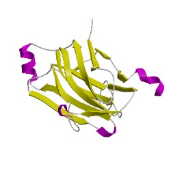
1qznA00
CATH Domain 1qznA00
PDB Chains (1)
| Chain ID | Date inserted into CATH | CATH Status |
| A | 05 Mar, 2006 | Chopped
|
CATH Domains (1)
UniProtKB Entries (1)
| Accession |
Gene ID |
Taxon |
Description |
| Q7WYN3 |
Q7WYN3_9FIRM |
Hungateiclostridium cellulolyticum |
Cellulosomal scaffoldin adaptor protein B |