×
Network disruptions
We have been experiencing disruptions on our local network which has affected the stability of these web pages.
We have been working with IT support team to get this fixed as a matter of urgency and apologise for any inconvenience.
PDB Information
| PDB | 1HEF |
| Method | X-RAY DIFFRACTION |
| Host Organism | Escherichia coli |
| Gene Source | Human immunodeficiency virus 1 |
| Primary Citation |
The crystal structures at 2.2-A resolution of hydroxyethylene-based inhibitors bound to human immunodeficiency virus type 1 protease show that the inhibitors are present in two distinct orientations.
Murthy, K.H., Winborne, E.L., Minnich, M.D., Culp, J.S., Debouck, C.
J.Biol.Chem.
|
| Header | Hydrolase/Hydrolase Inhibitor |
| Released | 1992-09-21 |
| Resolution | 2.200 |
| CATH Insert Date | 05 Mar, 2006 |
PDB Images (4)
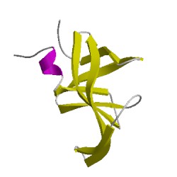
1hefE00
CATH Domain 1hefE00
PDB Chains (2)
| Chain ID | Date inserted into CATH | CATH Status |
| E | 05 Mar, 2006 | Chopped
|
| I | 05 Mar, 2006 | Rejected
|
CATH Domains (1)
UniProtKB Entries (1)
| Accession |
Gene ID |
Taxon |
Description |
| P03366 |
POL_HV1B1 |
Human immunodeficiency virus type 1 BH10 |
Gag-Pol polyprotein |