×
Network disruptions
We have been experiencing disruptions on our local network which has affected the stability of these web pages.
We have been working with IT support team to get this fixed as a matter of urgency and apologise for any inconvenience.
PDB Information
| PDB | 1BIB |
| Method | X-RAY DIFFRACTION |
| Host Organism | |
| Gene Source | Escherichia coli |
| Primary Citation |
Escherichia coli biotin holoenzyme synthetase/bio repressor crystal structure delineates the biotin- and DNA-binding domains.
Wilson, K.P., Shewchuk, L.M., Brennan, R.G., Otsuka, A.J., Matthews, B.W.
Proc.Natl.Acad.Sci.USA
|
| Header | Transcription Regulation |
| Released | 1992-07-06 |
| Resolution | 2.800 |
| CATH Insert Date | 05 Mar, 2006 |
PDB Images (5)
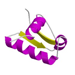
1bibA01
CATH Domain 1bibA01
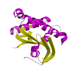
1bibA02
CATH Domain 1bibA02
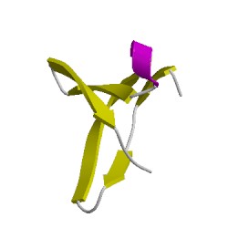
1bibA03
CATH Domain 1bibA03
PDB Chains (1)
| Chain ID | Date inserted into CATH | CATH Status |
| A | 05 Mar, 2006 | Chopped
|
CATH Domains (3)
UniProtKB Entries (1)
| Accession |
Gene ID |
Taxon |
Description |
| P06709 |
BIRA_ECOLI |
Escherichia coli K-12 |
Bifunctional ligase/repressor BirA |