×
Network disruptions
We have been experiencing disruptions on our local network which has affected the stability of these web pages.
We have been working with IT support team to get this fixed as a matter of urgency and apologise for any inconvenience.
PDB Information
| PDB | 2A5X |
| Method | X-RAY DIFFRACTION |
| Host Organism | |
| Gene Source | |
| Primary Citation |
The crystal structure of a cross-linked actin dimer suggests a detailed molecular interface in F-actin
Kudryashov, D.S., Sawaya, M.R., Adisetiyo, H., Norcross, T., Hegyi, G., Reisler, E., Yeates, T.O.
Proc.Natl.Acad.Sci.Usa
|
| Header | Contractile Protein |
| Released | 2005-07-01 |
| Resolution | 2.490 |
| CATH Insert Date | 05 Mar, 2006 |
PDB Images (5)
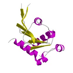
2a5xA01
CATH Domain 2a5xA01
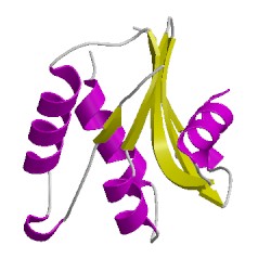
2a5xA02
CATH Domain 2a5xA02
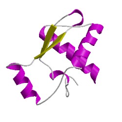
2a5xA03
CATH Domain 2a5xA03
PDB Chains (1)
| Chain ID | Date inserted into CATH | CATH Status |
| A | 05 Mar, 2006 | Chopped
|
CATH Domains (3)
UniProtKB Entries (1)
| Accession |
Gene ID |
Taxon |
Description |
| P68135 |
ACTS_RABIT |
Oryctolagus cuniculus |
Actin, alpha skeletal muscle |